• Radformation to Accelerate AI-Based Solutions with Limbus AI Acquisition

    April 30, 2024 – Radformation, Inc, a leader in automation solutions for cancer care announced today that it has acquired privately-held Limbus AI, a leading global provider of automated contouring software for radiation therapy. This acquisition signifies Radformation’s commitment to driving innovation and expanding its AI-driven capabilities in cancer care.

  • Read More
  • Limbus Contour

    Automatic Contouring for Radiation Therapy

    Clinical grade contours backed by comprehensive research.

    Install on your existing computers. Patient data stays local and secure.

    Request Demo arrow_forward_ios See Contours
  • Clinically validated deep learning segmentation for radiotherapy

    Fast

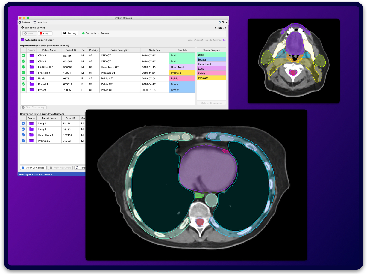
    Scans are contoured in seconds using preferred structure set templates from planning.

    Automated

    Contours generated automatically and sent immediately to treatment planning system after each scan. Configure once, implement in workflow forever.

    Turnkey

    Extensive library of clinically validated anatomical structures ready for immediate use out of the box.

    Secure

    State of the art deep learning entirely on local computers. No cloud transfer of patient data required.

  • Limbus Contour

Full Automation

CT images processed immediately after each scan. Limbus Contour detects and applies the correct clinical template, and sends the contours to the treatment planning system for manual review.

Limbus Contour Software Automation
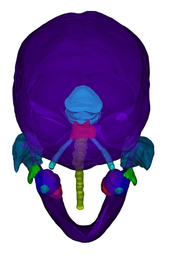
  • 3D Head Neck Segmentation
  • Easy to Setup

    AI contours up and running in minutes with your existing treatment planning system.

    • toggle_on

      Setup in Minutes

      Get automatic contouring up and running in less than an hour. Create Templates and customize contour metadata to match your treatment protocols. Configure Limbus Contour to detect the correct treatment site and apply the appropriate Template for planning.

    • personal_video

      Vendor Neutral

      Contours exported as compliant DICOM RT-Structure files. Supports a wide variety of treatment planning systems. No plug-ins or extensions needed.

    • done_all

      Supported Platforms

      Windows 10 and newer. No GPU required. One click install on an existing workstation. No cloud, no patient data privacy concerns.

Unmatched Security and Speed

Limbus Contour is the only autocontouring solution that runs entirely on your existing clinical workstations. No GPU or cloud data sharing required. Contours available in 1-3 minutes when running on a typical Intel CPU. GPU acceleration available for high-throughput use cases for even faster local contouring times.

  • Under 3 minutes

    Average contouring time on local CPU installations

  • 1 minute

    Average contouring time on local GPU installations

*Based on over 100,000 scans contoured by clinics worldwide

Expert Grade Contours

Select Scan view_list

5 - 10x faster than manual contouring

Extensive Structure Library

Limbus Contour features a large selection of organs at risk (OAR) and clincial target volume (CTV) contours that work out of the box immediately. No atlas creation or local curation of data necessary. All typical organs at risk and CTV structures included (260+ structures).

  • Head & NeckCT


    OARs

    Body

    Bone_Hyoid

    Bone_Mandible

    BrachialPlex_L

    BrachialPlex_R

    BrachialPlexs

    Brain

    Brainstem

    Cavity_Oral

    Cerebellum

    Clavicle_L

    Clavicle_R

    Cochlea_L

    Cochlea_R

    Cornea_L

    Cornea_R

    Esophagus

    Eye_L

    Eye_L

    Eyes

    Glnd_Lacrimal_L

    Glnd_Lacrimal_R

    Glnd_Submand_L

    Glnd_Submand_R

    Glnd_Thyroid

    Hippocampus_L

    Hippocampus_R

    InternalAuditoryCanal_L

    InternalAuditoryCanal_R

    Larynx

    Lens_L

    Lens_R

    Lips

    Lobe_Temporal_L

    Lobe_Temporal_R

    Musc_Constrict

    Musc_Sclmast_L

    Musc_Sclmast_R

    OpticChiasm

    OpticNrv_L

    OpticNrv_R

    Optics

    Parotid_L

    Parotid_R

    Pituitary

    Retina_L

    Retina_R

    Skin

    SpinalCanal

    SpinalCord

    CTVs

    LN_Neck_L

    LN_Neck_R

    LN_Neck_IA

    LN_Neck_IA6

    LN_Neck_IB_L

    LN_Neck_IB_R

    LN_Neck_II_L

    LN_Neck_II_R

    LN_Neck_III_L

    LN_Neck_III_R

    LN_Neck_IV_L

    LN_Neck_IV_R

    LN_Neck_V_L

    LN_Neck_V_R

    LN_Neck_VI

    LN_Neck_VIIA_L

    LN_Neck_VIIA_R

    LN_Neck_VIIB_L

    LN_Neck_VIIB_R

    LN_Neck_VIIAB_L

    LN_Neck_VIIAB_R

    LN_Neck_234_L

    LN_Neck_234_R

    LN_Neck_2347AB_L

    LN_Neck_2347AB_R

    CNSMR T1 Contrast


    OARs

    Brainstem

    Cornea_L

    Cornea_R

    Eye_L

    Eye_R

    Hippocampus_L

    Hippocampus_R

    Optics

    Retina_L

    Retina_R

    Gyne HDRCT


    OARs

    Bladder

    Bowel

    Canal_Anal

    Colon_Sigmoid

    Rectum

    Urethra

    Cylinder

    Gyne HDRMR


    OARs

    Bladder

    Bowel

    Canal_Anal

    Colon_Sigmoid

    Rectum

    Urethra

  • Thorax & AbdomenCT


    OARs

    A_Aorta

    A_Aorta_Base

    A_Aorta_I

    A_Celiac

    A_LAD

    A_Mesenteric_S

    A_Pulmonary

    Atrium_L

    Atrium_R

    Body

    Bowel_Bag

    Bowel_Bag_Superior

    Bowel

    Bowel_Superior

    BrachialPlex_R

    BrachialPlexs

    Breasts

    Breast_L

    Breast_R

    Breast_Implant_L

    Breast_Implant_R

    Bronchus

    Carina

    Chestwall

    Chestwall_L

    Chestwall_R

    CW2cm_L

    CW2cm_R

    Clavicle_L

    Clavicle_R

    Duodenum

    Esophagus

    Gallbladder

    Glnd_Thyroid

    GreatVes

    Heart

    Heart+A_Pulm

    Humerus_L

    Humerus_R

    Kidney_L

    Kidney_R

    Kidneys

    Liver

    Lung_L

    Lung_R

    Lungs

    Musc_PecMinor_L

    Musc_PecMinor_R

    Pancreas

    Pericardium

    Pericardium+A_Pulm

    Ribs

    Ribs_L

    Ribs_R

    Skin

    SpinalCanal

    SpinalCord

    Spleen

    Sternum

    Stomach

    Trachea

    V_Venacava_I

    V_Venacava_S

    VBs

    VB_C1

    VB_C2

    VB_C3

    VB_C4

    VB_C5

    VB_C6

    VB_C7

    VB_L1

    VB_L2

    VB_L3

    VB_L4

    VB_L5

    VB_T01

    VB_T02

    VB_T03

    VB_T04

    VB_T05

    VB_T06

    VB_T07

    VB_T08

    VB_T09

    VB_T10

    VB_T11

    VB_T12

    Ventricle_L

    Ventricle_R

    CTVs

    LN_Ax_L1_L

    LN_Ax_L1_R

    LN_Ax_L2_L

    LN_Ax_L2_R

    LN_Ax_L3_L

    LN_Ax_L3_R

    LN_Ax_Sclav_L

    LN_Ax_Sclav_R

    LN_IMN_L

    LN_IMN_R

    LN_IMN_L_Expand

    LN_IMN_R_Expand

    LN_Sclav_L

    LN_Sclav_R

    ESTRO_LN_Ax_IP_L

    ESTRO_LN_Ax_IP_R

    ESTRO_LN_Ax_L1_L

    ESTRO_LN_Ax_L1_R

    ESTRO_LN_Ax_L2_L

    ESTRO_LN_Ax_L2_R

    ESTRO_LN_Ax_L2+IP_Fill_L

    ESTRO_LN_Ax_L2+IP_Fill_R

    ESTRO_LN_Ax_L3_L

    ESTRO_LN_Ax_L3_R

    ESTRO_LN_IMN_L

    ESTRO_LN_IMN_R

    ESTRO_LN_IMN_L_Expand

    ESTRO_LN_IMN_R_Expand

    ESTRO_LN_Sclav_L

    ESTRO_LN_Sclav_R

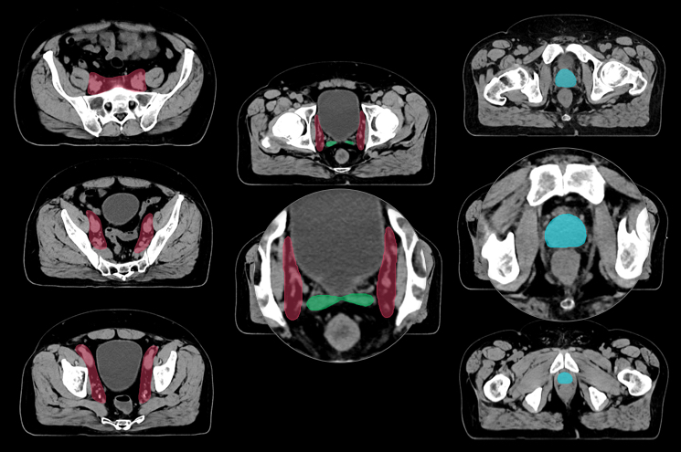
  • PelvisCT


    OARs

    Body

    Bladder

    Bone_Ilium_L

    Bone_Ilium_R

    Bone_Ilium

    Bone_Ischium_L

    Bone_Ischium_R

    Bone_Pelvic

    BoneMarrow_Pelvic

    Bowel

    Bowel_Extend

    Bowel_Full

    Bowel_Upper

    Bowel_Bag

    Bowel_Bag_Extend

    Bowel_Bag_Full

    Bowel_Bag_Upper

    Canal_Anal

    CaudaEquina

    Colon_Sigmoid

    Femur_Head_L

    Femur_Head_R

    Femur_Heads

    Mesorectum

    PenileBulb

    PubicSymphys

    Rectum

    Sacrum

    Skin

    Uterus+Cervix

    Vagina

    CTVs

    LN_Inguinal_L

    LN_Inguinal_R

    LN_Pelvics

    PelvisVessels

    Prostate

    Prostate+SeminalVes

    ProstateBed

    SeminalVes

    PelvisCBCT


    OARs

    Bladder

    Femur_Head_L

    Femur_Head_R

    Prostate

    Rectum

    SeminalVes

    CTVs

    LN_Pelvics

    PelvisMR T2


    OARs

    Bladder

    Femur_Head_L

    Femur_Head_R

    PenileBulb

    PubicSymphys

    Prostate

    Rectum

    Sacrum

    SeminalVes

    Combine structures to match your clinical protocols exactly.

    All structures work on full body and across different anatomic sites.

    Full list of structures not available in all markets.

    Contact Limbus AI for more information on availability in your region.

Clinical Target Volumes

Clinically validated CTVs to speed up the treatment planning process. Elective nodal CTVs across a variety of treatment sites. Configure Limbus Contour to export every 2nd, 3rd, or nth slice to speed up manual adjustments and review.

CTV nomenclature for structures refers to the tissue surrounding regions of interest for lymph nodes and other non-tumor target anatomy. Limbus Contour is not intended to automatically contour tumors or tumor clinical target volumes

  • Head / Neck
  • Breast
  • Pelvis
    • Head and Neck


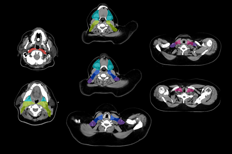
      Elective Neck Lymph Node CTV

      Choose from a combined structure volume including level IB, II, III, IV, V, VIIA, VIIB, VIIa/b. Or choose individual split sub-levels.

      • circleLN_Neck_IA

        circleLN_Neck_IA6

        circleLN_Neck_IB

        circleLN_Neck_II

        circleLN_Neck_III

        circleLN_Neck_IV

        circleLN_Neck_V

        circleLN_Neck_VI

        circleLN_Neck_VIIA

        circleLN_Neck_VIIB

        circleLN_Neck_VIIAB

        circleLN_Neck_234

        circleLN_Neck_2347AB

        circleLN_Neck

    • CTV Head Neck
    • Breast


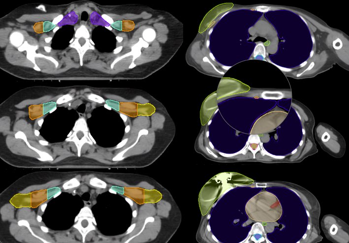
      Breast Regional Lymph Node CTVs

      Choose from a combined structure volume including Axillary levels I, II, III and supraclavicular lymph node regions. Or choose individual split sub-levels.

      • circleLN_Ax_L1

        circleLN_Ax_L2

        circleLN_Ax_L3

        circleLN_Sclav

        circleLN_Ax_Sclav

      Internal Mammary Lymph Node CTV (IMC)

      Spanning the first three intercostal spaces.

      • circleLN_IMN

      ESTRO style Breast Lymph Node volumes included

    • CTV Breast
    • Pelvis


      Pelvis Lymph Node CTV

      Pelvis lymph node region spanning lymph nodes including obturator lymph nodes, internal and external iliac lymph nodes (cropped anteriorly at level of femoral heads) up to common iliac lymph nodes (L5/S1 region).

      • circleLN_Pelvics

        circleLN_Inguinal

        circlePelvisVessels

      Prostate and SV

      Prostate and seminal vesicle to speed up manual prostate CTV contouring.

      • circleProstate

        circleSeminalVes

        circleProstate + SeminalVes

        circleProstateBed

    • CTV Pelvis

    Continuously Improving

  • playlist_add_check

    Expert Datasets

    Segmentation models are trained on large datasets which have been meticulously reviewed by experts and standardized to meet contouring guidelines.

  • local_hospital

    Clinically Driven

    External testing and continuous prospective clinical evaluation ensures each organ model is reliable.

  • refresh

    Better with Time

    AI contours undergo routine clinical evaluation and re-training to fine tune and improve models over time. New structures are added regularly.

    Models are subject to regulatory clearance and may not be available in all markets.

What our users are saying

Tens of thousands of treatments have been planned using Limbus Contour. Here’s what clinicians have to say:

  • "We explored various auto-contouring solutions and found that Limbus Contour was the only product liked by our dosimetrists. It integrated seamlessly into our existing workflow, representing the closest to 'plug-n-play' medical software that I have encountered. Because the software runs on a local server it greatly simplified the approval process with our hospital's IT department. Limbus Contour has enhanced our productivity and our ability to manage urgent planning requests, making them much more manageable.”

    Greg Salomons, PhD - Medical Physicist & Assistant Professor

    Dept. of Oncology - Cancer Centre of Southeastern Ontario (CCSEO) - Queen’s University

  • "After testing various systems, we decided on Limbus AI a few months ago. After several months of use in clinical routine, we have never regretted our decision to go with Limbus AI and we no longer want to do without it.”

    Dr. Kay Willborn - Head of Radiation Oncology

    Pius Hospital Oldenburg

  • "Limbus has reduced the time spent contouring in our dosimetrists and physicians by 75% and 55% respectively. We are subsequently starting our IMRT and SBRT patients days earlier than planned while providing dosimetry more time to focus on planning and optimizing. The ease of use, speed and accuracy has made Limbus AI the most indispensable software tool in our clinic.”

    Joe Dise - Landauer Medical Physicist

    Missouri Baptist

  • "Having been a Limbus AI user for several years, I am very glad to be able to continue this exciting experience after recently having moved to a different facility. My colleagues and I appreciate the smart workflow and the enormous amount of time saved – and the high quality of the contours generated by Limbus AI."

    Stefan Höcht - Vice Director

    Klinikum Ernst von Bergmann, Potsdam, Germany

  • "We were one of the first users of Limbus Contour in Germany and are very happy with it. It is fully integrated in our workflow and saves time in the treatment planning process. With Limbus Contour, auto-contouring of organs at risk as well as nodal regions is very efficient and effective and rarely needs manual adaptations. This young and dynamic company keeps its contouring according to international standards up to date.”

    Dr. Silke Tribius

    Asklepios Clinic - Hamburg, Germany

  • "We aim to deliver radiotherapy treatments in the shortest timeframe possible. At the same time, radiotherapy is evolving rapidly with Adaptive treatment, Hypofractionation, and SABR which require very accurate structure definition and thus thinner slices. This increases the required time for contouring, placing more pressure on staffing resources and workflow. AI based algorithms offer great potential to reduce this pressure.

    From our initial investigation, Limbus Contour is providing excellent results for multiple sites. For example, within minutes we can create the main contours required for a prostate treatment. After reviewing numerous cases we have found that the contours produced required very minimal editing. This allows us to very significantly decrease the amount of time required before starting the planning stage.

    The software is very easy to setup and straightforward to use. We have also found the Limbus team extremely responsive to our requests with excellent support, even though we are very far away and in a completely different time zone."

    Jerome Gastaldo - Chief Physicist

    St. Georges Cancer Center

Research

Performance backed by leading research. Limbus AI is committed to proving the value of AI in medicine through published peer reviewed research.

Cancers (Open Access)

December 2023New

Clinical Use of a Commercial Artificial Intelligence-Based Software for Autocontouring in Radiation Therapy: Geometric Performance and Dosimetric Impact

"Our results show that the combination of automatically generated contours from Limbus Contour™ and careful review by a clinical radiation oncologist results in time saving without affecting the quality of treatment plan. In conclusion, after quality checks that involve both geometric accuracy as well as dosimetric impact, contouring based on AI can be safely adopted in clinical practice."

View Publicationlibrary_books

Artificial Intelligence in Medicine

October 2023New

Artificial Intelligence–Based Autosegmentation: Advantages in Delineation, Absorbed Dose-Distribution, and Logistics

"In great extent, AI (i.e. Limbus Contour v.1.6.0) yielded clinically acceptable OARs and certain clinical target volumes in the explored anatomic segments. Sparse correction and assessment requirements place AI+C (i.e. AI and Radiation Oncologist) as a standard workflow. Minimal clinically relevant differences in OAR exposure were identified. A substantial amount of person-hours could be repurposed with this technology."

View Publicationlibrary_books

Brachytherapy

December 2022

Evaluation of Autocontouring in the Interstitial HDR Brachytherapy of Liver Tumors

"The application of artificial intelligence to interstitial HDR brachytherapy of the liver brachytherapy can shorten the treatment by about 15 minutes on average. This reduces the unpleasant lying time with applicators for the patient."

View Abstractlibrary_books

Life

December 13, 2022

Implementation of a Commercial Deep Learning-Based Auto Segmentation Software in Radiotherapy: Evaluation of Effectiveness and Impact on Workflow

"Automatic contouring was able to significantly reduce the time required in the procedure, simplifying the workflow, and reducing interobserver variability. Its implementation was able to improve the radiation therapy workflow in our department."

View Publicationlibrary_books

Radiotherapy & Oncology (Green Journal)

Sept 01, 2022

A Randomized Blinded Assessment of a Machine Learning Based Autocontouring Tool for Breast Cancer Radiotherapy Compared to Peer-Reviewed Radiation Oncologist Contours

View Publicationlibrary_books

March 01, 2020

Comparing deep learning-based auto-segmentation of organs at risk and clinical target volumes to expert inter-observer variability in radiotherapy planning

View Publicationlibrary_books

International Journal of Environmental Research and Public Health

April 11, 2022

Clinical Validation of a Deep-Learning Segmentation Software in Head and Neck: An Early Analysis in a Developing Radiation Oncology Center

"Our results suggest that AC could become a useful time-saving tool to optimize workload and resources in RT departments."

View Publicationlibrary_books

British Institute of Radiology - AI in Practice 2022 Meeting

March 17, 2022

Qualitative Evaluation of Auto-segmented Structures for Radiotherapy Planning Using Peer Review Guidelines

"Reviewers found that 70.3% of the structure sets required none, or “minor” modification. Looking at the OaRs in isolation, then the percentage of structure sets requiring “minor” or no modification rises to 95.2%."

View Publicationlibrary_books

Radiation Oncology (Open Access Journal)

December 2023New

Integrating Artificial Intelligence Into Radiation Oncology: Can Humans Spot AI?

"The results indicate that AI can perform radiation contouring comparably to human oncologists but much faster. The challenge faced by professionals in identifying AI versus human contours highlights AI's advanced capabilities in medical tasks."

View Publicationlibrary_books

June 08, 2021

Implementation of Deep Learning-Based Auto-Segmentation for Radiotherapy Planning Structures: A Multi-Center Workflow Study

Recipient of CARO 2020 Award for Best Abstract in Science and Applied Technology

View Publicationlibrary_books

Frontiers in Oncology

June 07, 2021

Training and Validation of Deep Learning-Based Auto-Segmentation Models for Lung Stereotactic Ablative Radiotherapy Using Retrospective Radiotherapy Planning Contours

View Publicationlibrary_books

Practical Radiation Oncology

June 27, 2020

Clinical evaluation of deep learning and atlas based auto-contouring of bladder and rectum for prostate radiotherapy

View Publicationlibrary_books

ESTRO 2023

The impact of LIMBUS Al based contouring on the efficiency of prostate radiotherapy planning

“Our results have shown that editing a Limbus auto-contour to a clinical standard was more time efficient than manually contouring structures for prostate plans, with a significant time saving benefit. DSC values showed good agreement between AIC and EC from the Limbus system. For all structures, substantial time was saved editing an AIC, when compared to generating a new EC. Our study shows that Al can safely be used as a substantial time saver in the prostate radiotherapy planning process.”

View Abstractlibrary_books

ASTRO 2020 and CARO 2020

  • Applications of Deep Learning for Automatic Contouring of Tumors in the Brain

    Recipient of CARO 2020 Award for Abstracts Submitted by the Medical Student Research and Mentorship Awardees

  • Deep Learning-based Auto-Segmentation for Pelvic Organs at Risk and Clinical Target Volumes in Intracavitary High Dose Rate Brachytherapy

    View Abstractlibrary_books

ASTRO 2019

  • Comparing Deep Learning-based Auto-segmentation of Organs at Risk and Clinical Target Volumes to Expert Inter-Observer Variability in Radiotherapy Planning

    View Abstractlibrary_books
  • Validation of Deep Learning-based Auto-Segmentation for Organs at Risk and Gross Tumor Volumes in Lung Stereotactic Body Radiotherapy

    View Abstractlibrary_books
  • Automatic Deep Learning-based Segmentation of Brain Metastasis on MPRAGE MR Images for Stereotactic Radiotherapy Planning

    View Abstractlibrary_books